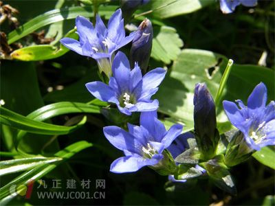
Method of identification of gentian: (1) Cross-section of this product: gentian epidermal cells sometimes remain and the outer wall is thick. The cortex is narrow; the cells in the outer cortex are square, the wall is slightly thick, and the cork is corked; the endothelial cells are tangentially elongated, and each cell is divided into several square-like small cells by the longitudinal wall. The phloem is wide and has cracks. The formation layer is not very obvious. 3 to 10 clusters of xylem vessels. The pith is obvious. The parenchyma cells contain fine calcium oxalate needles. The tissue outside the endothelial layer of the gentian has been shed. The xylem tube is well developed and evenly covered. No pith. The powder is yellowish brown. The surface of the gentian outer cortex cells is spindle-shaped, and each cell is divided into several small square cells by a transverse wall. The surface of the endothelial cells is rectangular, very large, and the lateral wall is slender and laterally textured. Each cell is divided into several small cells by the mediastinum wall, and most of the mediastinum wall is thickened by beads. The parenchyma cells contain fine calcium oxalate needles. The mesh and ladder conduits have a diameter of approximately 45 μm. The gentian has no outer cortical cells. The endothelial cells are square or rectangular, and the lateral texture of the flat wall is thick and dense, and some are as thick as 3 μm. Each cell is divided into a plurality of grid-shaped small cells, and the partition wall is slightly thickened or beaded. (2) Take 0.5 g of the powder of the product, add 5 ml of methanol, immerse for 4 to 5 hours, filter, and concentrate the filtrate to about 2 ml as a test solution. Another gentamicin reference substance was added, and methanol was added to make a solution containing 2 mg per 1 ml as a reference solution. According to the thin layer chromatography (Appendix VIB) test, 5 μl of each of the above two solutions was taken and placed on the same silica gel GF254 thin layer plate with sodium carboxymethyl cellulose as a binder, with ethyl acetate-methanol-water ( 20:2:1) as a developing agent, secondly unrolled, taken out, dried, and placed under ultraviolet light (254 nm) for inspection. In the chromatogram of the test sample, spots of the same color are displayed at positions corresponding to the chromatogram of the reference substance. Microscope Accessories ,Microscope Eyepiece,Microscope Camera Adapter,Microscope Slides And Cover Slips Ningbo Beilun Kalinu Optoelectronic Technology Co.,Ltd , https://www.yxmicroscope.com